Pelvic Floor Laxity With Cystocele

What Is Pelvic Organ Prolapse And What You Can Do About It Pelvic Organ Prolapse Urinary Incontinence Incontinence
Anterior Vaginal Repair Your Pelvic Floor
Your Guide To The Pelvic Floor Nuffield Health

Perineal Descent Austin Urogynecology

Pelvic Floor Exercises Getting To Know Your Pelvic Floor Mutu System
Prolapse Pelvic Floor Series 3 Foundational Concepts
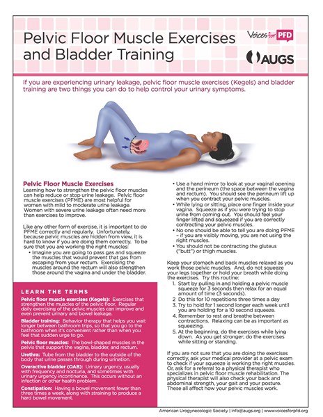
Biofeedback or weighted vaginal cones may also help the patient with low degrees of cystocele to strengthen the pelvic floor.
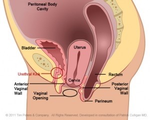
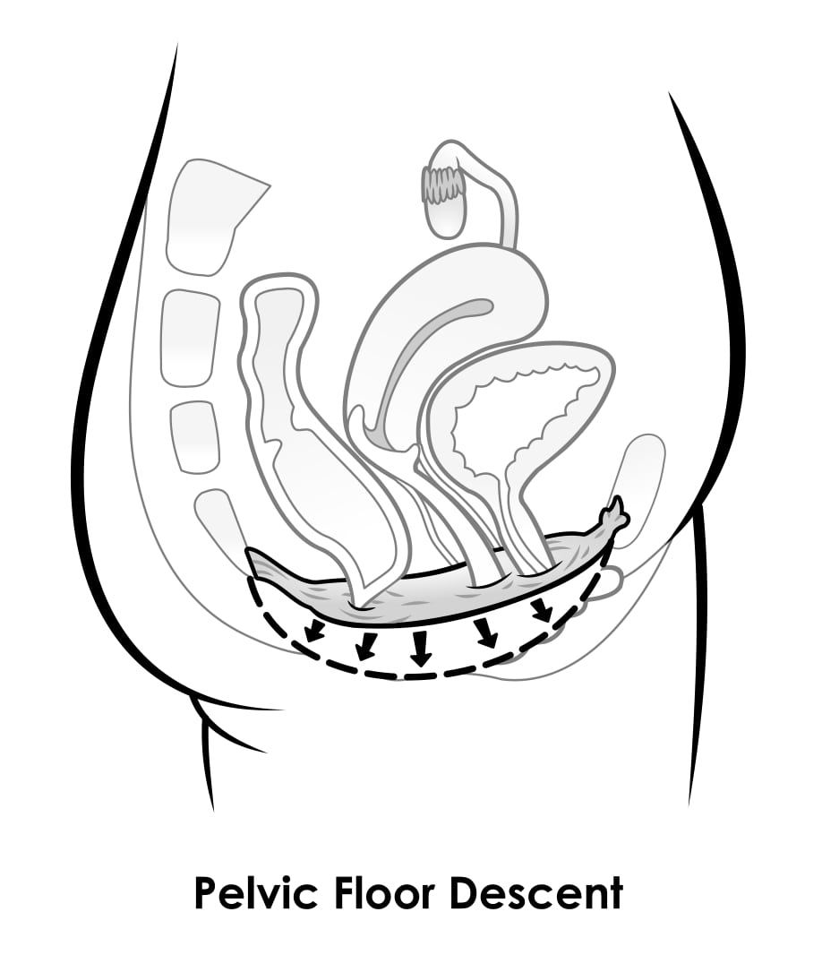
Pelvic floor laxity with cystocele. Up to 50 of such women have some degree of pelvic prolapse. More specifically the term anterior repair refers to correction of the front wall of the vagina. Similarly the study by fielding et al. A rectocele may be an isolated finding or occur as part of a generalized weakening of the pelvic floor muscles.
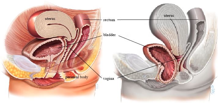
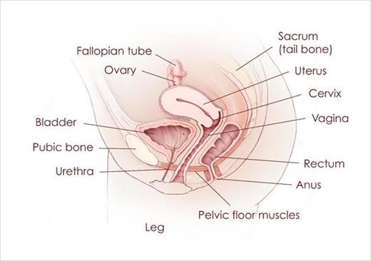
Pelvic floor weakness abnormal descent of the bladder cystocele uterus or vagina uterine or vaginal vault prolapse small bowel enterocele or rectum rectocele is a significant women s health problem that primarily affects parous women over 50 years of age. Pelvic floor repair the most common surgery for prolapse is a pelvic floor repair which is a broad term used to describe simple surgical repairs of the pelvic floor. And posterior repair refers to correction of the back wall of the vagina. Hold the contraction for five seconds and then relax for five seconds.
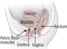
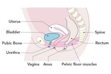
A prolapse of the front anterior wall of the vagina is usually due to a weakness in the strong tissue layer fascia that divides the vagina from the bladder. A woman with such device in place can observe by means of the analog. A cystocele also called a prolapsed or dropped bladder is the bulging or dropping of the bladder into the vagina. Tighten contract your pelvic floor muscles the muscles you use to stop urinating.
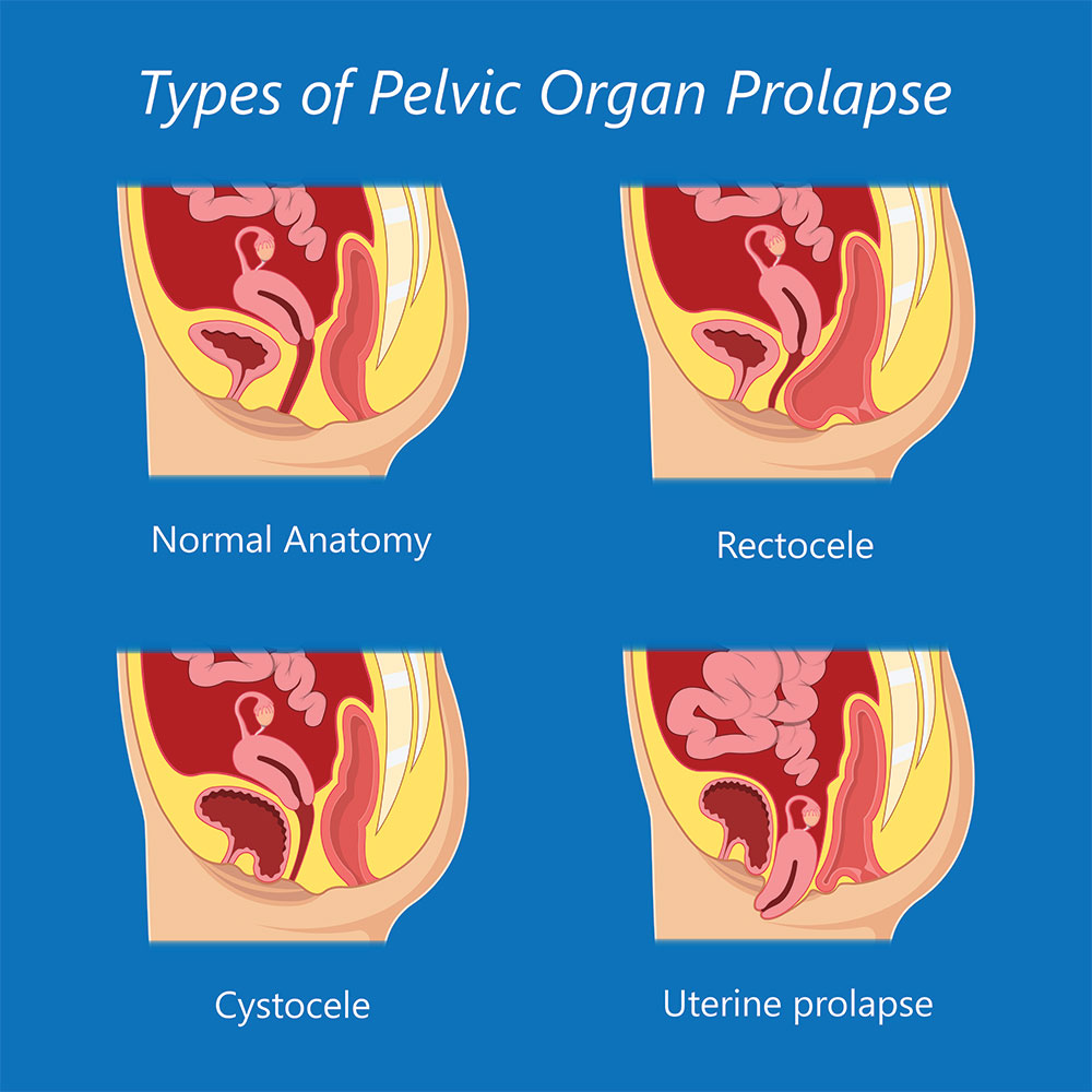
About 1 in 10 women who have had children require surgery for vaginal prolapse. In fact about one third of all women are affected by prolapse or similar conditions over their lifetime. Other pelvic organs such as the bladder cystocele and the small intestine enterocele can bulge into the vagina leading to similar symptoms as rectocele. Cystocele mri pelvic floor dysfunction rectocele.
If this is too difficult start by holding for two seconds and relaxing for three seconds work up to holding the contraction for 10 seconds at a time. Showed that although a greater degree of pelvic floor laxity was shown on mri in the sitting position it was not superior to standard supine mri. Pelvic organ prolapse a type of pelvic floor disorder can affect many women. The bladder located in the pelvis between the pelvic bones is a hollow muscular balloon shaped organ that expands as it fills with urine.
Moreover in a 0 5 t open mri system one must contend with images of a lower snr and soft.
Physical Therapy Treatments Voices For Pfd

Weightlifting After Cystocele Repair Pelvic Floor Article Hystersisters Pelvic Floor Pelvic Floor Exercises Safe Exercises

Pelvic Floor Therapy Optimaliving Therapy Wellness

Illustration Of The Female Pelvic Floor Showing Relationships Of The Download Scientific Diagram
Pelvic Organ Prolapse My Doctor Online
Prolapse And Running Physio Guide To Avoid Prolapse Worsening

Pelvic Floor Pt The Transgender Experience Body Connect Health And Wellness
Treating Pelvic Organ Prolapse Harvard Health
Prolapse Bladder Weakness Jean Hailes

Uterine Prolapse Uterus Obg Uterine Prolapse Bladder Prolapse Prolapsed Uterus

Pelvic Organ Prolapse Pelvic Organ Prolapse Pelvic Congestion Syndrome Uterine Prolapse

Pelvic Floor Dysfunction Gastrointestinal Society
Https Www Ics Org Workshops Handoutfiles 000197 Pdf

