Pie In The Floor Defect
Visual Field Defects Ophthalmology Flashcards Memorang
Optic Radiation Lesions Pie In The Sky Pie On The Floor Youtube

Quadrantanopia An Overview Sciencedirect Topics
Pupillary Pathway Amp Field Defects Dr K Srikanth 25 05 16

Pie In The Sky Vs Pie On The Ground Usmle Forum
Visual Pathway Neurology Medbullets Step 1
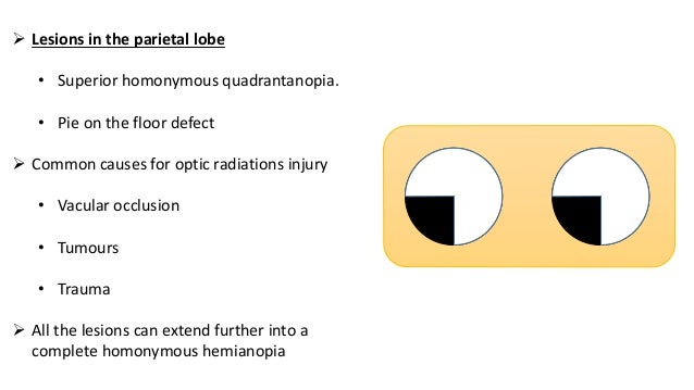
Ischemia or bleeding from a stroke is the most common cause for a quadranopsia.
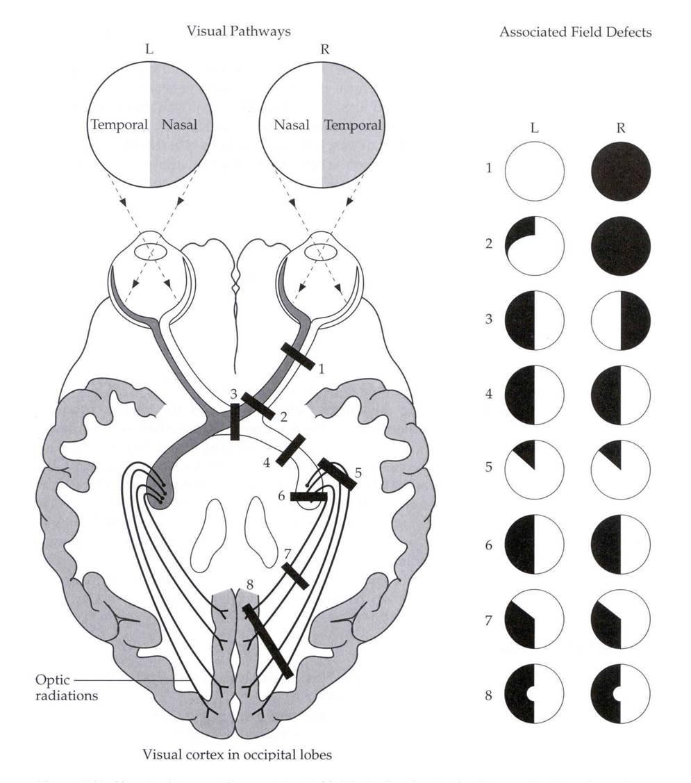
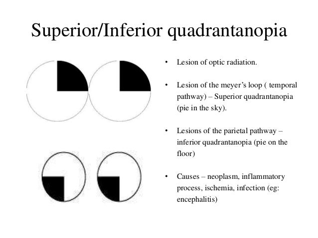
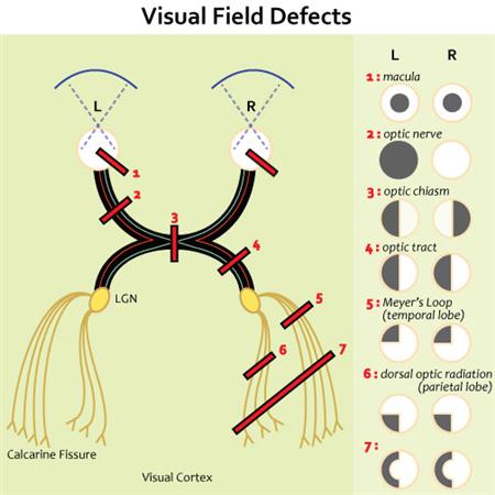
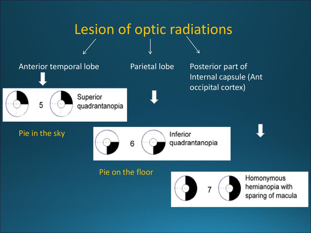
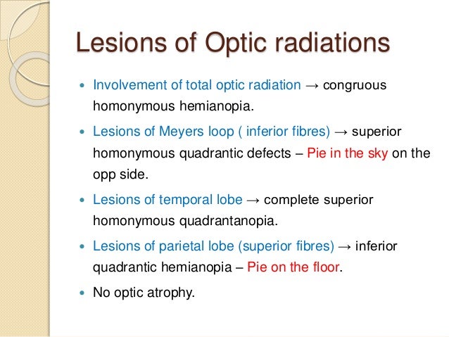
Pie in the floor defect. Lesions at a particular site of the visual pathway result in a specific visual field defect depicted on the right hand side 500 reprinted from australian family physician vol. Superior fibers inferior field defect course directly posteriorly from the lgb into the parietal lobe. Quadrantanopia quadrantanopsia refers to an anopia affecting a quarter of the field of vision. Optic radiation lesions pie in the sky pie on the floor.
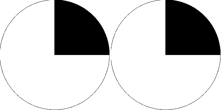
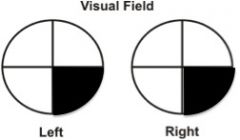
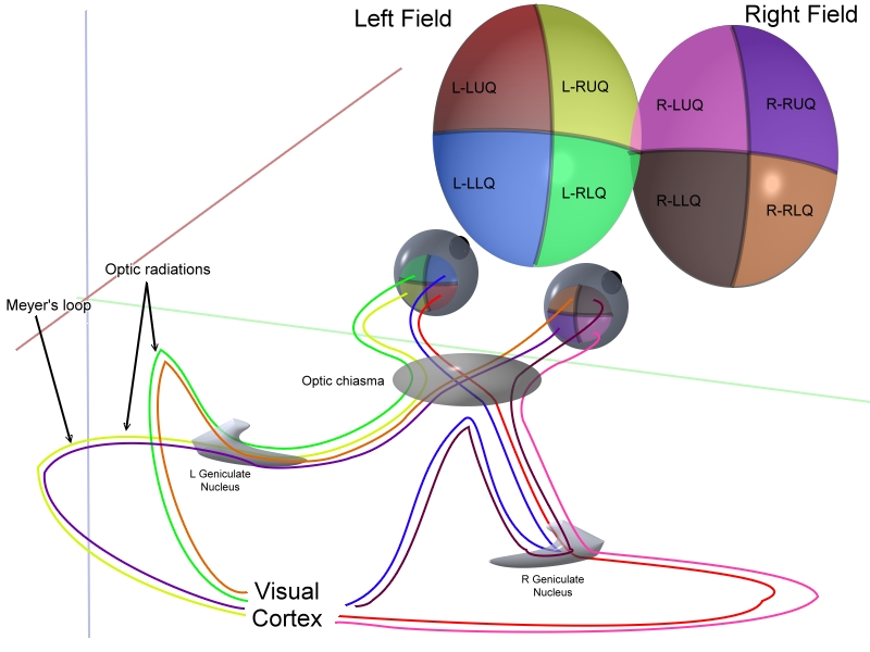
Scotoma ipsilateral optic nerve. Neuroanatomy of the visual pathway and the blood supply. While quadrantanopia can be caused by lesions in the temporal and parietal lobes it is most commonly associated with lesions in the occipital lobe. A superior homonymous loss of vision in two quarters due to a lesion of the most anterior and inferior fibres of the optic radiations in the temporal lobe involving meyer s loop on the contralateral side of the visual pathway fig.
Cardio69 07 13 13 15 28. A superior quadranopsia or pie in the sky defect points to a contralateral temporal lobe problem whereas an inferior quadranopsia pie on the floor points to the contralateral parietal lobe. It can be associated with a lesion of an optic radiation. Re visual field defect question 2912251.
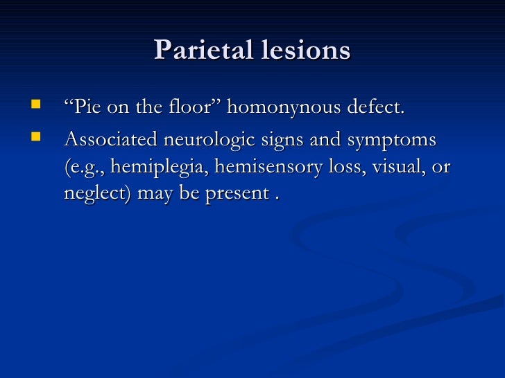
Decerberate and decorticate posturing. Pie in the floor defect this set is often in folders with. Parietal lesions pie on the floor homonynous defect. Homonymous congruous or incongruous pie shaped defects below the horizontal meridian.
Anopia or central scotoma ipsilateral optic chiasm 2. Understanding of the visual system is paramount 1. Anterior temporal lobe pie on the sky homonymous. I see pie in the floor.
Brain stem umn and descending lmn tracts. Anterior at junction with one of the optic nerves. Start studying visual field defects fancy names. Learn vocabulary terms and more with flashcards games and other study tools.
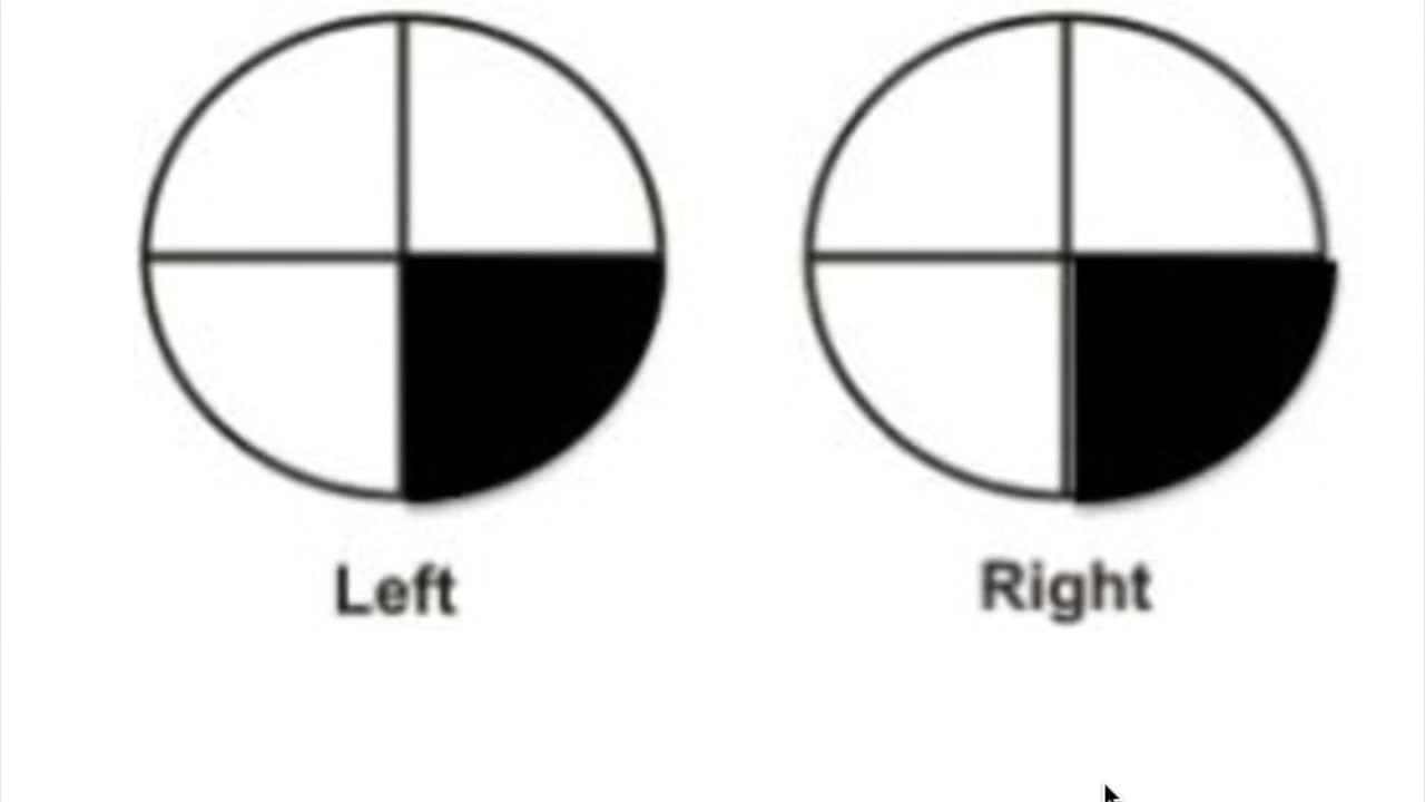
Pie on the floor. Visual pathway or visual field deficits are defects in visual space determined by the location of a lesion in the neurological visual pathway from eye to brain cortex. Associated neurologic signs and symptoms e g hemiplegia hemisensory loss visual or neglect may be present. E inferior quadrantanopia pie on the floor figure 2.
What would be the diff in occipital lobe lesion parietal lobe lesion regard to homonymous quadrantanopia pat in both lesion will say doc.
Quadrantanopia Wikipedia
Anatomy And Lesions Of Visual Pathways

Common Visual Field Defects A Constriction Of The Visual Field B Ring Scotoma C Central Scotoma D Cecocen Infographic Health Medical Students Optometry
All Lesions To Visual Pathways Ssom Flashcards Cram Com
Visual Pathway
Gale Onefile Health And Medicine Document Visual Field Defects In Neurological Diseases
Optic Radiation Mediconotebook

Man Experiences Decreased Peripheral Vision

Visual Fields Flashcards Quizlet

An Intro To Neuro Optometry Optometry School Neuro
Visual Pathway And Lesion

Optic Radiation Wikiwand
Visual Pathway Lesions Ppt Download

