Pelvic Floor Ultrasound Technique

Instrumentation And Techniques For Perineal And Introital Pelvic Floor Ultrasound Abdominal Key

Pelvic Floor Ultrasound Radiology Key
Instrumentation And Techniques For Translabial And Transperineal Pelvic Floor Ultrasound Radiology Key

Pdf Investigation Of Transabdominal Real Time Ultrasound To Visualise The Muscles Of The Pelvic Floor Semantic Scholar

Ultrasound Imaging Of The Pelvic Floor Part Ii Three Dimensional Or Volume Imaging Dietz 2004 Ultrasound In Obstetrics Amp Gynecology Wiley Online Library

Standard Acquisition Screen Of 3d Pelvic Floor Ultrasound When Using Download Scientific Diagram

Relaxation techniques such as yoga and stretching can also help to relax your pelvic floor muscles.
Pelvic floor ultrasound technique. A computer converts the. Because translabial ultrasound is the most commonly used modality for pelvic floor evaluation it is the focus of this chapter. Most slings are highly echogenic and therefore their type and mode of action are easily visualised on an ultrasound. There are techniques described in literature that use translabial ultrasound i e transducer is placed on the labia majora.
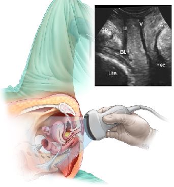
A modification of the translabial or transperineal technique is introital imaging which typically uses high frequency endocavitary transducers placed in the introitus. The exam normally involves two components. This is a presentation by dr andrew edwards. The technique is similar to 2d imaging with orientation set in the midsagittal plane.
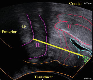
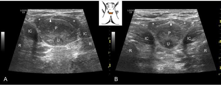
Transperineal ultrasound refers to ultrasound performed with the transducer position on the perineum. To obtain volumes an acquisition angle between 70 and 85 is needed to capture pertinent anatomy levator hiatus vagina paravaginal tissue urethra puborectalis muscle anorectal angle. Taking warm baths is another useful technique. Physiotherapist stuart turner demonstrates how real time ultrasound can be used to assess and retrain the muscles of the pelvic floor.
The intended audience are gp s and gynaecologists. Hans dietz gives an overview of different techniques of imaging in urogynecology followed by a detailed discussion of pelvic floor ultrasound to assess anatomy and function of the urethra and bladder. It describes the ultrasound technique and use of pelvic floor. A transabdominal ta evaluation and a transvaginal tv endovaginal ev evaluation.
These sound waves bounce off your organs and tissues and then echo back to the transducer. Warm water improves blood circulation and relaxes. Pelvic ultrasound is usually the initial modality for imaging gynecologic pathology including acute pelvic pain and chronic pelvic pain. Term introital ultrasound 10 is also used where transducer is placed at the vaginal introitus or posterior fourchette.
Transperineal Pelvic Floor Ultrasound Scan Your Pelvic Floor
Https Www Iuga Org Files 55 Pelvic Floor Imaging 14 Pelvic Floor Ultrasound Basic Settings And Procedures V2018 Pdf
Musculoskeletal Sonography And Occupational Performance Laboratory Msop

Pelvic Floor Ultrasound An Underutilised But Useful Diagnostic Tool Express Healthcare

Pelvic Floor Ultrasound Mutuaterrassa Youtube

Novel Insight Into The Dynamics Of Male Pelvic Floor Contractions Through Transperineal Ultrasound Imaging Sciencedirect

Ultrasound Imaging Of The Pelvic Floor Part I Two Dimensional Aspects Dietz 2004 Ultrasound In Obstetrics Amp Gynecology Wiley Online Library

Https Encrypted Tbn0 Gstatic Com Images Q Tbn 3aand9gcqpkxf1csczfi Beaudjlshcbwlecqflzo18q Usqp Cau
Transperineal Ultrasound Tpus An Exciting New Technology Women S And Men S Health Physiotherapy
Perineal Pelvic Floor Ultrasound Applications And Literature Review Abdominal Key

Pelvic Floor Assessment By Ultrasound Youtube
Https Www Jospt Org Doi Pdf 10 2519 Jospt 2007 2548
Herman Wallace Transabdominal Ultrasound In The Assessment Of Abdominal And Pelvic Floor Muscles

