Pelvic Floor Laxity Radiology

Is Ligament Laxity Running Your Life Too Breathe Pelvic Floor Guest Posting

Pin On Anatomy

Female Bladder Leakage Solutions To Get Control Ucla Obstetrics Gy Ucla Health Oncology Nursing

Research Lumbosacral Transition Vertebrae Ltv Luw In The Dutch Shepherd Institute Of Genetics Shepherd Animal Hospital Vertebrae

Pin On Things I Wrote
Https Pubs Rsna Org Doi Pdf 10 1148 Rg 345140137
Symptoms such as obstructive defecation incontinence and sphincter complex disorders have a significant impact on patient lifestyle and physical mental well being 1 2.
Pelvic floor laxity radiology. Muscles of the pelvic floor can be imaged with mri and pelvic floor movements can be assessed with dynamic mri. Mr imaging of the pelvic floor. Pelvic floor dysfunction is a common condition where you re unable to correctly relax and coordinate the muscles in your pelvic floor to urinate or to have a bowel movement. El sayed rf el mashed s farag a morsy mm abdel azim ms.
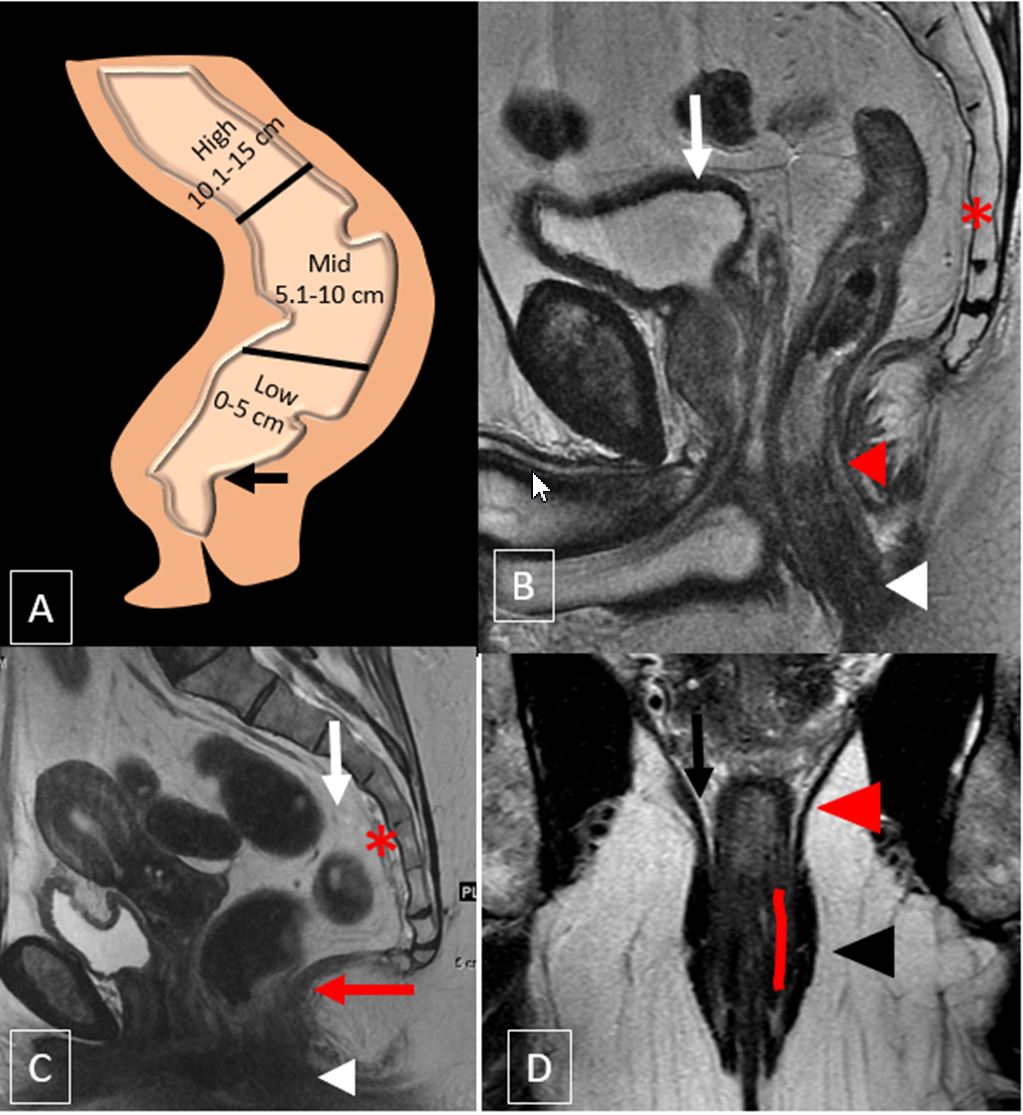
Imaging studies include colonic transit to assess bowel motility. Dynamic cystoproctography is still the reference imaging technique for assessing functional pelvic organ. Provides limited visualization of the pelvic muscles. Imaging is required to assess pelvic floor laxity in women with urinary and bowel continence issues.
There are four phases of evaluation. Our objectives are to 1 increase awareness of sui in young females 2 test our hypothesis with an upright vcug and 3 report. Clinical scanners have platforms identical to dedicated research scanners enabling rapid clinical translation. Compared sitting mr defecography with dynamic supine mri and showed that sitting mr defecography is not superior to dynamic supine mri for depiction of clinically relevant.
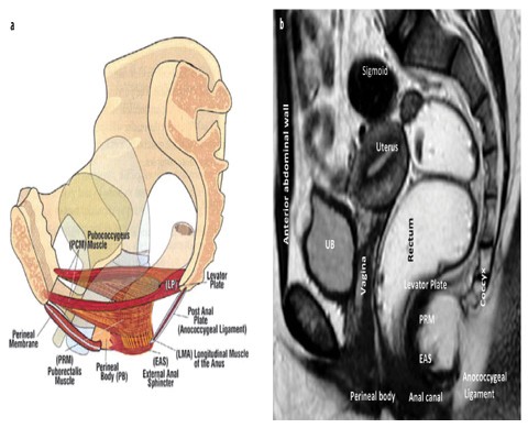
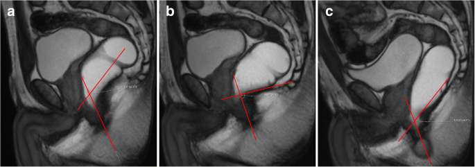
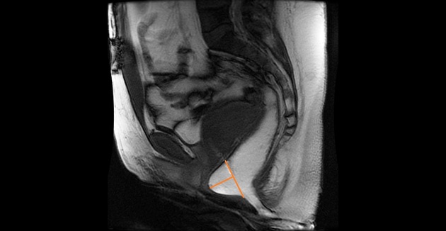
On these images the radiologist identifies the pubococcygeal line which represents the level of the pelvic floor the h and m lines which are helpful for confirming pelvic floor laxity and the angle of the levator plate with the pubococcygeal line which is helpful for identifying small bowel prolapse. Assessment with combined analysis of static and dynamic mr imaging findings. Pelvic floor imaging is an important part of both gastrointestinal and functional urology urogynaecological departments. Physically active adolescent females develop pelvic floor laxity demonstrable on upright vcug.
Dynamic imaging imaging obtained at rest during squeezing straining and defecation has a central role in the diagnosis of pelvic floor dysfunction and it is crucial when choosing a conservative versus a surgical treatment 9. Body mri research at stanford is fostered by a tight link between research scientists in the department of radiology the university and throughout the bay area. These efforts are supported by multiple nih grants. Seynaeve r billiet i vossaert p verleyen p steegmans a.
If you re a woman you may also feel pain during sex and if you re a man you may have problems having or keeping an erection erectile dysfunction or ed. Stress urinary incontinence sui is common among older multiparous females but rarely reported in active young girls.

Pin On Famous Buttocks
How To Perform And Report Magnetic Resonance Imaging For Pelvic Floor Dysfunction An Interactive Case Based Approach Springerlink
Https Www Abdominalradiology Org Resource Resmgr Education Dfp Pelvicfloor Mri Imaging Mri Of Pelvic Floor Dysfunct Pdf

Types Of Incontinence Explained Nursing School Survival Nursing School Notes Nursing School Tips

Pelvic Floor Laxity Body Mri

Pin On Famous Buttocks

Pin By Great Wolf On Radius Fractures Fractures Dislocation

Peroneal Tendon Syndromes Ankle Anatomy Ankle Ligaments Torn Ligament In Ankle
Dynamic Magnetic Resonance Imaging Of The Female Pelvic Floor A Pictorial Review Springerlink

Assessment Of Pelvic Organ Prolapse A Review Shek 2016 Ultrasound In Obstetrics Amp Gynecology Wiley Online Library
Epos
Anterior Rectocoele Radiology Case Radiopaedia Org

