Pelvic Floor Injury On A Ct Scan

Why A Doctor Would Order A Ct Of The Abdomen And Pelvis Diagnostic Imaging Services
Preoperative Pelvic Ct Scan Of The Patient The Arrow Indicates Primary Download Scientific Diagram

A Pelvic Computed Tomography Ct Scan Shows A High Density Shadow In Download Scientific Diagram

Ct Abdomen Pelvis Revealing A Moderate Amount Of Free Fluid In The Download Scientific Diagram

Postoperative Pelvic Pain An Imaging Approach Sciencedirect

Mri Defaecating Proctogram The Xray Doctor Mri Health Articles Mens Health

Ct scans continue to be an essential tool in the management of trauma especially with regards to pelvic injuries.
Pelvic floor injury on a ct scan. A ct scan uses x rays to look at bones muscles body organs and blood vessels. When are they useful. Ct is the modality of choice for accurately depicting complex acetabular or pelvic ring fractures. Your doctor may order a ct scan to examine a fracture pattern or assess the extent of damage in the hip joint.
Pelvis and proximal femora. A ct computed tomography scan uses x rays to make detailed pictures of your body and structures inside your body. A pelvic ct scan takes pictures of your pelvis the area between your hips. Pelvic incidence is the angle subtended by a line connecting the femoral heads to a perpendicular bisector of the s1 endplates.
Pelvic incidence was measured on ct scan with sagittal reconstructions using specific ct slices. These muscles look like a hammock or sling stretched from the tailbone at the back to the pubic bone in front and from one sitting bone to the other. Who they can help. Pelvic fractures carry a significant risk of uncontrolled pelvic bleeding and exsanguination from pelvic fractures is a real possibility.
What can these scans do. Your pelvic floor is the group of muscles and ligaments in your pelvic region the pelvic floor acts like a. During the test you will lie on a table that is attached to the ct scanner. Pelvic floor dysfunction is the inability to control the muscles of your pelvic floor.
If your doctor recommends a pelvis ct scan you likely have questions. The increased availability and improvements in imaging techniques has confirmed ct scans as an important tool in the detection of life threatening injuries resulting in a marked reduction in the overall number of pelvic fractures missed. The pelvic incidence was measured on a ct scan by a new method described below. A ct scan of the pelvis can give the doctor information about the bones and organs in the pelvic area.
The pelvic floor consists of the muscles and tissues that support the pelvic organs including the uterus bladder bowel and rectum in women and the bladder bowel rectum and prostate in men. Each picture also called a slice shows a few layers of your body tissue on a computer or tv like screen. Your caregiver may do a ct scan. A ct scan uses x rays and a computer to create two and three dimensional pictures of the hip and pelvic bones enabling doctors to examine a fracture from many different angles.
The ct scanner is a large doughnut shaped machine.
A Pelvic Ct Scan Shows Calcified Sling Tape Arrow Eroding The Bladder Download Scientific Diagram

Anti N Methyl D Aspartate Nmda Receptor Encephalitis With Frustrated Diagnosis Course A Case Report Nmda Receptor Rare Disease Neurology

Retroaortic Left Renal Vein An Incidental Finding On Approximately 3 Of Ct Scans Important To Report To Inform Surgery Www Thexray Ct Scan Renal Radiology

Ps59037ds Jpg Doctor Stock Ct Scan Radiology Spine Health

Pin On Tailbone Pain Doctor
Https Pubs Rsna Org Doi Pdf 10 1148 Rg 241035063
Chronic Pelvic Pain Following Childbirth Orthopedic Trauma

Pelvis Ct Congenital Spine Deformity Spina Bifida In A Patient Paralysis Radiologist Radiol Diagnostic Imaging Protected Health Information Radiology

Pelvis Ct Scan Shows A Hip Dislocation With Fracture In A Patient After Car Accident Radiologis Hip Dislocation Bones And Muscles Diagnostic Imaging

Pin On Tailbone Mri

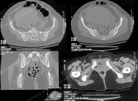
Pelvis Computed Tomograph Axial Ct Ct Scan Scan Pelvis

Pin On Mri

Crash Ct Scan Guidance In 2020 Ct Scan Mri Brain Scan

